Home
Uncategories
Back Of Neck Anatomy - Pin En Tips To Help Relieve Back Pain : Contains glands ( thyroid, parathyroid, and thymus ), the larynx, pharynx and trachea.
Back Of Neck Anatomy - Pin En Tips To Help Relieve Back Pain : Contains glands ( thyroid, parathyroid, and thymus ), the larynx, pharynx and trachea.
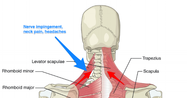
Back Of Neck Anatomy - Pin En Tips To Help Relieve Back Pain : Contains glands ( thyroid, parathyroid, and thymus ), the larynx, pharynx and trachea.. The muscles of the back muscles make up a large part of the anatomy (structure) of the back. The muscles of the neck run from the base of the skull to the upper back and work together to bend the head and assist in breathing. The human neck is one of the most complex structures we have because it contains many important elements that converge in a very small space. Neck anatomy pictures bones muscles nerves. Located at the back and side of the neck, the levator scapulae muscle connects the neck's cervical spine with the shoulder.
The hard white exterior covering of the tooth is the enamel. The inner portions of the tooth consist of the dentin, a bonelike tissue, and the pulp. Contain the common carotid artery, internal. The superficial lymph nodes of the head and neck receive lymph from the scalp, face and neck. Each nerve provides sensation to a specific area of the body called a dermatome.
The Link Between Posture And Chronic Neck And Upper Back Pain Back Pain And Headache Specialist Burke Va Nova Headache Chiropractic Center from images.squarespace-cdn.com The skull is a strong, bony capsule that rests on the neck and encloses the brain. In the back, the neck reaches the c7 vertebra. The inner portions of the tooth consist of the dentin, a bonelike tissue, and the pulp. Located at the back and side of the neck, the levator scapulae muscle connects the neck's cervical spine with the shoulder. The neck region, or cervical region when referring to its spinal location, is a crucial area of the body as it contains nervous pathways connecting the brain to the peripheral nerves and is vital in head mobility, which aids in the body's sensing of its environment. Back pain is common and might be caused by a problem with a muscle. These two ligaments connect and support the spine from the neck to the lower. An area called the occiput.
The neck is one of the most complex and intricate structures in our body and includes the spinal cord, which sends messages from the brain to the rest of the body.
The neurocranium (cranial vault) and the viscerocranium (facial skeleton). They ultimately drain into the deep lymph nodes. The muscles of the neck are present in four main groups. The neck muscles, including the sternocleidomastoid and the trapezius, are responsible for the gross motor movement in the muscular system of the head and neck. Located at the back and side of the neck, the levator scapulae muscle connects the neck's cervical spine with the shoulder. The occipital bone is the only bone in your head that connects with your cervical spine (neck). The muscles of the neck run from the base of the skull to the upper back and work together to bend the head and assist in breathing. The larynx is located where the pharynx, the back of the mouth and nasal cavity, divides into the trachea (the tube that carries air to the lungs) and the esophagus (the tube that carries food to. The motion of the muscles of the neck are divided into four. The skull is a strong, bony capsule that rests on the neck and encloses the brain. Two of the main ligaments in the back are the anterior longitudinal ligament and the posterior longitudinal ligament. Choose from 500 different sets of flashcards about neck anatomy back neck upper on quizlet. Anatomy of back of human neck, anatomy of the back and neck, anatomy of the back of the neck, anatomy of the back of the neck muscles, anatomy of the back of your.
The rotation function takes the head into the opposite side to which this neck and shoulder muscle is located. These two ligaments connect and support the spine from the neck to the lower. The hard white exterior covering of the tooth is the enamel. In particular, the levator scapulae muscle is susceptible to injury. It runs down the back part of the neck, and opens into the external jugular vein just below the middle of its course.
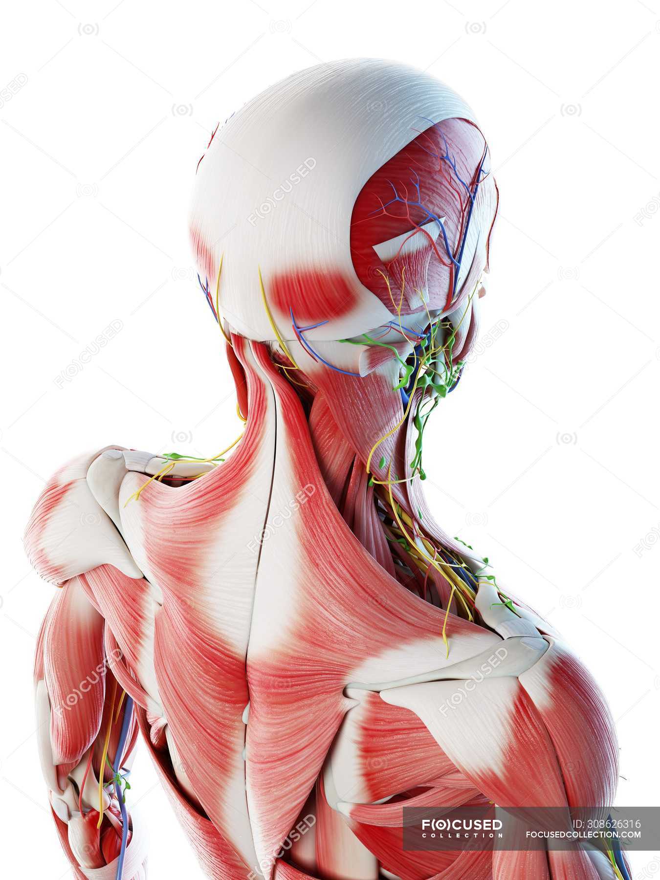
Male Back Neck And Head Muscles Computer Illustration Anatomical 3d Model Stock Photo 308626316 from st.focusedcollection.com Neck anatomy explained the neck begins at the base of the skull and connects to the thoracic spine (the upper back). Back pain is common and might be caused by a problem with a muscle. Working in pairs on the left and right sides of the body, these muscles. By far the most common cause of a stiff neck is a muscle strain or soft tissue sprain. Openstax chapter 11 for bio 201 by mattpearcy issuu. Then it extends to the clavicles and the sternum in front. Find symptoms,causes and treatments of back pain.for your health. This muscle is controlled by the third and fourth cervical.
Anatomy of back of human neck, anatomy of the back and neck, anatomy of the back of the neck, anatomy of the back of the neck muscles, anatomy of the back of your.
The occipital bone is a bone that covers the back of your head; Think of it like a jigsaw puzzle, all the pieces fit in together and are required to get the full picture as to how it works. Neck anatomy pictures bones muscles nerves. It also helps extend, tilt, and rotate your neck, which has the effect of bringing your head back, to the side, and turning it. The neck is essentially a passageway for air, food, liquids, blood, and more to travel between the head and the rest of the body, through structures such as blood vessels, nerves, and lymph nodes, as well as the larynx, trachea, and esophagus. The neck is the area between the skull base and the clavicles. These two ligaments connect and support the spine from the neck to the lower. Back pain is common and might be caused by a problem with a muscle. The skull is a strong, bony capsule that rests on the neck and encloses the brain. Working in pairs on the left and right sides of the body, these muscles. Anatomy of back of human neck, anatomy of the back and neck, anatomy of the back of the neck, anatomy of the back of the neck muscles, anatomy of the back of your. Contains cervical vertebrae and postural muscles. The rounded upper projections of the back teeth are cusps.
Muscle head anatomy vocal organ diagram female neck anatomy neck wireframe head neck human anatomy head artery anatomy face pharynx vector neck degree head anatomy 3d. As the tooth tapers below the gumline, the neck is formed. They ultimately drain into the deep lymph nodes. The neck is connected to the upper back through a series of seven vertebral segments. The neck is connected to the upper back through a series of seven vertebral segments.
Male Back Neck And Head Muscles Computer Illustration Anatomical 3d Model Stock Photo 308626316 from st.focusedcollection.com The cervical spine, your neck, is a complex structure making up the first region of the spinal column starting immediately below the skull and ending at the first thoracic vertebra. Openstax chapter 11 for bio 201 by mattpearcy issuu. See anatomy of the head and neck stock video clips. The hard white exterior covering of the tooth is the enamel. Neck anatomy nerves picture there are 8 spinal nerves that originate from the cervical spine. Muscle head anatomy vocal organ diagram female neck anatomy neck wireframe head neck human anatomy head artery anatomy face pharynx vector neck degree head anatomy 3d. It consists of two major parts: The majority of these nerves control the functions of the upper extremities and allow you to feel your arms, shoulder, and back of your head.
The neck is connected to the upper back through a series of seven vertebral segments.
The occipital bone is the only bone in your head that connects with your cervical spine (neck). Choose from 500 different sets of flashcards about neck anatomy back neck upper on quizlet. They are arranged in a ring shape; Back pain is common and might be caused by a problem with a muscle. These two ligaments connect and support the spine from the neck to the lower. They move the head in every direction, pulling the skull and jaw towards the shoulders, spine, and scapula. Contain the common carotid artery, internal. The occipital bone surrounds a large opening known as the foramen magnum. Working in pairs on the left and right sides of the body, these muscles. The cervical spine, your neck, is a complex structure making up the first region of the spinal column starting immediately below the skull and ending at the first thoracic vertebra. Neck anatomy explained the neck begins at the base of the skull and connects to the thoracic spine the upper back. Each nerve provides sensation to a specific area of the body called a dermatome. Below the neck, holding the tooth into the bone, is the root of the tooth.
0 Comments:
Post a Comment